- Electric and Telecom Plans Free
- Fire and Emergency Plans Free
- Floor Plans Free
- Plant Layout Plans Free
- School and Training Plans Free
- Seating Plans Free
- Security and Access Plans Free
- Site Plans Free
- Sport Field Plans Free
- Business Process Diagrams Free
- Business Process Mapping Free
- Classic Business Process Modeling Free
- Cross-Functional Flowcharts Free
- Event-driven Process Chain Diagrams Free
- IDEF Business Process Diagrams Free
- Logistics Flow Charts Free
- Workflow Diagrams Free
- ConceptDraw Dashboard for Facebook Free
- Mind Map Exchange Free
- MindTweet Free
- Note Exchange Free
- Project Exchange Free
- Social Media Response Free
- Active Directory Diagrams Free
- AWS Architecture Diagrams Free
- Azure Architecture Free
- Cisco Network Diagrams Free
- Cisco Networking Free
- Cloud Computing Diagrams Free
- Computer Network Diagrams Free
- Google Cloud Platform Free
- Interactive Voice Response Diagrams Free
- Network Layout Floor Plans Free
- Network Security Diagrams Free
- Rack Diagrams Free
- Telecommunication Network Diagrams Free
- Vehicular Networking Free
- Wireless Networks Free
- Comparison Dashboard Free
- Composition Dashboard Free
- Correlation Dashboard Free
- Frequency Distribution Dashboard Free
- Meter Dashboard Free
- Spatial Dashboard Free
- Status Dashboard Free
- Time Series Dashboard Free
- Basic Circle-Spoke Diagrams Free
- Basic Circular Arrows Diagrams Free
- Basic Venn Diagrams Free
- Block Diagrams Free
- Concept Maps Free
- Family Tree Free
- Flowcharts Free
- Basic Area Charts Free
- Basic Bar Graphs Free
- Basic Divided Bar Diagrams Free
- Basic Histograms Free
- Basic Line Graphs Free
- Basic Picture Graphs Free
- Basic Pie Charts Free
- Basic Scatter Diagrams Free
- Aerospace and Transport Free
- Artwork Free
- Audio, Video, Media Free
- Business and Finance Free
- Computers and Communications Free
- Holiday Free
- Manufacturing and Maintenance Free
- Nature Free
- People Free
- Presentation Clipart Free
- Safety and Security Free
- Analog Electronics Free
- Audio and Video Connectors Free
- Basic Circuit Diagrams Free
- Chemical and Process Engineering Free
- Digital Electronics Free
- Electrical Engineering Free
- Electron Tube Circuits Free
- Electronic Block Diagrams Free
- Fault Tree Analysis Diagrams Free
- GHS Hazard Pictograms Free
- Home Automation and Wiring Free
- Mechanical Engineering Free
- One-line Diagrams Free
- Power Сircuits Free
- Specification and Description Language (SDL) Free
- Telecom and AV Circuits Free
- Transport Hazard Pictograms Free
- Data-driven Infographics Free
- Pictorial Infographics Free
- Spatial Infographics Free
- Typography Infographics Free
- Calendars Free
- Decision Making Free
- Enterprise Architecture Diagrams Free
- Fishbone Diagrams Free
- Organizational Charts Free
- Plan-Do-Check-Act (PDCA) Free
- Seven Management and Planning Tools Free
- SWOT and TOWS Matrix Diagrams Free
- Timeline Diagrams Free
- Australia Map Free
- Continent Maps Free
- Directional Maps Free
- Germany Map Free
- Metro Map Free
- UK Map Free
- USA Maps Free
- Customer Journey Mapping Free
- Marketing Diagrams Free
- Matrices Free
- Pyramid Diagrams Free
- Sales Dashboard Free
- Sales Flowcharts Free
- Target and Circular Diagrams Free
- Cash Flow Reports Free
- Current Activities Reports Free
- Custom Excel Report Free
- Knowledge Reports Free
- MINDMAP Reports Free
- Overview Reports Free
- PM Agile Free
- PM Dashboards Free
- PM Docs Free
- PM Easy Free
- PM Meetings Free
- PM Planning Free
- PM Presentations Free
- PM Response Free
- Resource Usage Reports Free
- Visual Reports Free
- House of Quality Free
- Quality Mind Map Free
- Total Quality Management TQM Diagrams Free
- Value Stream Mapping Free
- Astronomy Free
- Biology Free
- Chemistry Free
- Language Learning Free
- Mathematics Free
- Physics Free
- Piano Sheet Music Free
- Android User Interface Free
- Class Hierarchy Tree Free
- Data Flow Diagrams (DFD) Free
- DOM Tree Free
- Entity-Relationship Diagram (ERD) Free
- EXPRESS-G data Modeling Diagram Free
- IDEF0 Diagrams Free
- iPhone User Interface Free
- Jackson Structured Programming (JSP) Diagrams Free
- macOS User Interface Free
- Object-Role Modeling (ORM) Diagrams Free
- Rapid UML Free
- SYSML Free
- Website Wireframe Free
- Windows 10 User Interface Free
Medical Mycology
Mycology is a branch of biology that studies fungi, their structure, properties, main characteristics and ways of infection. It is closely related to medicine, dermatology, toxicology, immunology, allergology and other fields. Fungi are incredibly widespread, and they come in all kinds, shapes, and sizes. However, there are both useful and destructive species for humans.
Fungi cause disease in humans, animals, and plants, spoil food, and can destroy entire crops. At the same time, they are natural decomposers, break down organic materials and play a decisive role in the environment. Fungi also promote plant growth because they convert the nitrogen present in the atmosphere into a form usable by plants. Some fungi are used as "useful" in food, agricultural, alcohol and other industries.
Medical mycology is a branch of mycology dealing with fungal infections. Pathogenic fungi and invasive fungal infections affect billions of people and animals worldwide. Therefore, they are under the close supervision of medical scientists who study their features, reactions, modifications, mutations and human susceptibility. This knowledge is driving progress in the diagnosis and treatment of fungi, and drug development.
Fungi pose a serious threat to human health. They cause a complex set of disease states, skin infections, allergies, infections of the mucous membranes, diseases of individual organs or systemic lesions of the body, inflammation, affect nails, hair, lungs. Fungal infections are most dangerous for people with weakened immune systems, with malignant tumors, HIV infection, diabetes mellitus, chronic diseases, violation of the protective bacterial microflora, after injuries, operations, organ transplants, in old age, etc. In severe cases, they can cause tissue destruction, organ failure and lethal cases.
In most cases, fungi are difficult to study because they are problematic or impossible to grow in the lab. In addition, the period of the fruiting body containing spores is short. However, medical mycology has firmly established itself in developed countries and its development continues permanently. Work on the classification of fungi, the development of diagnostic tests in the laboratory, epidemiological and environmental studies are ongoing and, in general, have a positive effect in the fight against fungal diseases and the development of effective methods of treatment.
The Medical Mycology solution for ConceptDraw DIAGRAM software provides a huge collection of vector design elements and a set of ready-made samples. These design tools are effective for compiling an overview of fungal diseases, their etiological agents, identifying the diversity of fungal pathogens, dimorphic fungi, mold, their role in systemic diseases, immunopathology, fungal mycotoxins. They help to describe the results of research in the field of medical mycology and immunology of fungi, the latest advances in the development of antifungal drugs. The solution is useful for demonstrating the concepts and methods for diagnosing fungal diseases and treating mycosis, for illustrating the most dangerous effects of fungi on humans and animals, fungal meningitis. It helps to tell about personal hygiene and other ways to avoid infection, the importance of complex antifungal therapy and immunotherapy. The solution for medical mycology is useful for microbiologists, clinical laboratory scientists, researchers, medical scientists, professors of medicine, students and interns.
-
Buy this solution $49 -
Solution Requirements - This solution requires the following products to be installed:
ConceptDraw DIAGRAM v18 - This solution requires the following products to be installed:
-
Support for this Solution -
Helpdesk
The Medical Mycology solution contains 16 examples and 17 libraries containing 365 vector graphics and icons, to allow you to create professional-looking illustrations, diagrams, and infographics inspired by medical mycology and fungal immunology science.
Design Elements — Fungi Microscopy

Design Elements — Fungal Diseases

Design Elements — Fungi Biology

Design Elements — Fungi Culture

Design Elements — Antifungal

Design Elements — Cleaning and Disinfecting

Design Elements — Eye Mycoses

Design Elements — Fungal Meningitis

Design Elements — Fungi Hosts

Design Elements — Mold

Design Elements — Mycoses Diagnosis

Design Elements — Mycoses Epidemiology

Design Elements — Mycoses Prevention

Design Elements — Mycoses Symptoms

Design Elements — Mycoses Transmission

Design Elements — Personal Hygiene

Design Elements — Skin and Nail Mycoses
Examples
There are a few samples that you see on this page which were created in the ConceptDraw DIAGRAM application by using the Medical Mycology solution. Some of the solution's capabilities as well as the professional results which you can achieve are all demonstrated here on this page.
All source documents are vector graphic documents which are always available for modifying, reviewing and/or converting to many different formats, such as MS PowerPoint, PDF file, MS Visio, and many other graphic ones from the ConceptDraw Solution Park or ConceptDraw STORE. The Medical Mycology solution is available to all ConceptDraw DIAGRAM users to get installed and used while working in the ConceptDraw DIAGRAM diagramming and drawing software.
Example 1: Antifungal Resistance in Aspergillus
This diagram was created in ConceptDraw DIAGRAM using a combination of libraries from the Medical Mycology Solution. An experienced user spent 10 minutes creating this sample.
This sample describes Aspergillus fumigatus. It is a fungus in the environment, which causes an invasive life-threatening infection "aspergillosis". The immunocompromised people with chronic diseases or earlier severe respiratory infections are the most susceptible. The infection occurs as a result of breathing fungi spores with air. Triazole antifungal drugs or azoles are the primary treatment for aspergillosis. They kill most of Aspergillus fumigatus in the body. However, according to research, 19% of Aspergillus fumigatus are resistant to azoles. This phenomenon is caused by the long-term treatment of patients chronically infected with Aspergillus fumigatus and complicates treatment. This also happens outside when azole fungicides are applied against Aspergillus fumigatus, harming plants and destroying harvests. Fungi develop the ability to survive, grow, multiply, and spread despite the exposition of antifungals. Any use of antifungals can cause the development of antifungal resistance, therefore the control of their responsible use and in some cases, replacement with alternative protective ways is extremely important.

Example 2: Blastomyces Life Cycle
This diagram was created in ConceptDraw DIAGRAM using a combination of libraries from the Medical Mycology Solution. An experienced user spent 10 minutes creating this sample.
This sample shows the life cycle of Blastomyces. These are fungi that cause blastomycosis in humans and animals. They live in wet soil, decaying and decomposing wood and leaves, shaded areas close to water sources like streams, rivers, and lakes. These fungi are spread in areas surrounding the Ohio and Mississippi River valleys, the Great Lakes, in Canada. Sometimes Blastomyces multiply in the garbage in damp premises, sheds, and basements. A common way of infection is through breathing the microscopic fungal spores from the air. Blastomyces live in the environment as mold. Once getting the organism, the spores grow as large-celled budding yeast in the lungs. The first symptoms include cough, fever, and fatigue. In common, treatment is simple with systemic antifungal drugs. However, the infection can spread through the bloodstream to other organs, the central nervous system, skin, bones, joints, and take serious forms if it is not treated. Muscle or joint aches, chest pain, weight loss occur between 3 weeks and 3 months after infection. There is no vaccine to prevent blastomycosis. However, getting spores into the body does not necessarily lead to the development of infection, the strong immune system helps to avoid negative effects.

Example 3: Coccidioides Life Cycle
This diagram was created in ConceptDraw DIAGRAM using a combination of libraries from the Medical Mycology Solution. An experienced user spent 10 minutes creating this sample.
This sample refers to coccidioidomycosis, or valley fever, caused by the fungi Coccidioides immitis and Coccidioides posadasii. These fungi are common in the southwestern United States, Mexico, Central and South America. Fungi spores are found in the soil as molds with hyphae partitions, which are easily fragmented into arthroconidia and spread through the air with dust. The infection enters the body from the air and microscopic fungal spores settle in the lungs. Here, coccidioid spores turn into large tissue-penetrating globules, grow and rupture, releasing thousands of small endospores and spreading into surrounding tissues. These endospores form new spherules. Coccidioidomycosis is a pulmonary and hematogenous disease. It can present with a variety of clinical manifestations, including fatigue, fever, cough, dyspnea, headache, muscle pain, and rash. Antifungal medications are effective against it. However, in most cases, the disease is asymptomatic, and people recover on their own within weeks or months.

Example 4: Control Mold
This diagram was created in ConceptDraw DIAGRAM using a combination of libraries from the Medical Mycology Solution. An experienced user spent 10 minutes creating this sample.
Mold is a type of fungi that grows and spreads on damp or decaying organic substances indoors or outdoors. These highly invasive microorganisms emit dusty clouds of toxic spores, harmful to people sensitive to mold, who have respiratory allergies, asthma, or hypersensitivity pneumonitis. As for buildings and homes, mold grows in wet places, on windows with condensation, pipes, tiles, and joints in premises with excessive moisture like bathrooms, damp attics, basements, and roofs. It is also found on wood and paper products, drywall, carpets, fabric, etc. The most common indoor molds include Penicillium, Aspergillus, and Cladosporium. The effective ways to control mold primarily include its removing from surfaces using bleach or other cleansers, without dangerous mixing them and at the obligatory wearing rubber gloves, goggles, or even boots to protect himself during cleanup. They also include the elimination of the sources of moisture, promptly fixing any leaks, cleaning and drying after flooding, ventilation premises, especially shower, laundry, and cooking areas, and controlling humidity using the air conditioner or dehumidifier.

Example 5: Cryptococcus Gattii Life Cycle
This diagram was created in ConceptDraw DIAGRAM using a combination of libraries from the Medical Mycology Solution. An experienced user spent 10 minutes creating this sample.
This sample shows a life cycle of Cryptococcus gattii. These are encapsulated yeast spread in trees and soil around them in tropical and subtropical areas, Australia, New Zealand, British Columbia, Canada, some parts of the US. Infection enters the body of humans or animals from air through breathing. Microscopic spores affect the lungs, travel through the bloodstream, and infect the central nervous system or other parts of the body. Cryptococcus gattii causes pulmonary cryptococcosis, pneumonia, basal meningitis, cerebral cryptococcosis, formation of nodules in the lungs or brain. The yeast cells grow thick capsules to protect themselves, divide and multiply by budding. The main symptoms include cough, fever, shortness of breath, chest pain, headache, neck pain, sensitivity to light, nausea, vomiting, seizures, weight loss. Sometimes, the combination of symptoms is very unusual. The fact that the infection is rare and difficult to diagnose also complicates the situation.

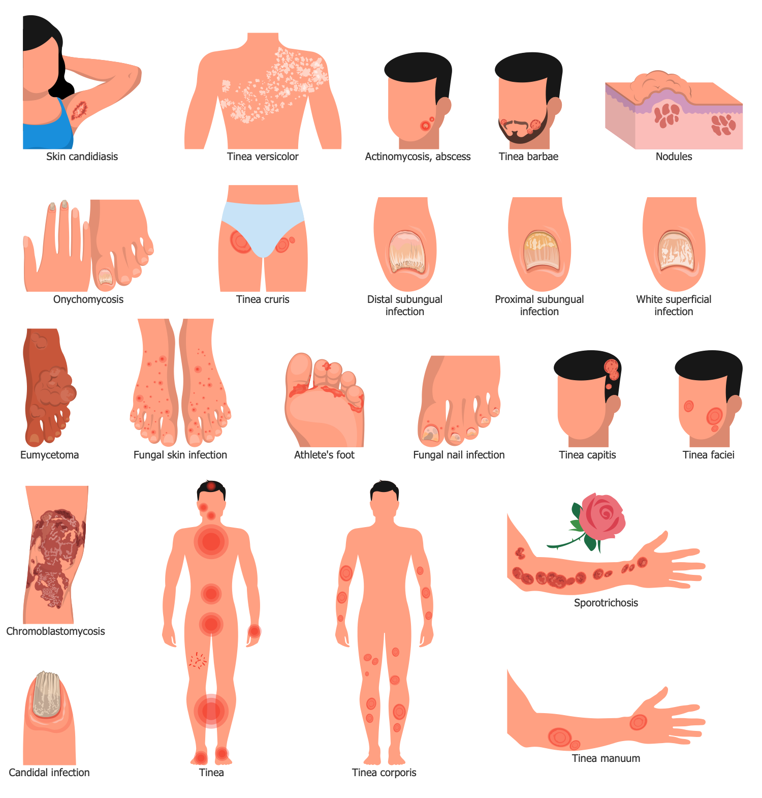
Example 6: Dermatophytosis Types
This diagram was created in ConceptDraw DIAGRAM using a combination of libraries from the Medical Mycology Solution. An experienced user spent 10 minutes creating this sample.
This sample is an overview of dermatophytosis, also known as ringworm or tinea. This is a fungal infection of the skin caused by about 40 types of fungi that feed on the protein keratin. Keratin is the structural material of the outer layer of human skin and nails. Therefore, dermatophytosis is localized on the skin of the body or scalp, feet, hands, in the beard area, nails. It is characterized by a red, itchy, scaly, and circular rash, and hair loss in the affected area. Risk factors include a weak immune system, contact with animals, contact sports, and walking barefoot in public places such as public showers. The incubation period is four to fourteen days after contact with the pathogen. However, with strong immunity, causative agents of dermatophytosis can settle on human skin and not cause infection. Pathogens are detected under a microscope when examining a culture of scrapings. Local treatment is carried out with antifungal creams (clotrimazole, miconazole). In severe cases, perioral antifungals are also prescribed.

Example 7: Fungus Cell Cycle
This diagram was created in ConceptDraw DIAGRAM using a combination of libraries from the Medical Mycology Solution. An experienced user spent 15 minutes creating this sample.
This sample shows the stages of the life cycle of a fungal cell from its "birth" as a result of the division of the mother cell to its reproduction and its own division into two daughter cells. The life cycle of eukaryotic cells includes two main phases: interphase and mitotic phase. Interphase is the phase of cell growth, copying of its genetic material (DNA), and preparation for mitosis. The mitotic (M) phase is the stage of cell division into two daughter cells, followed by the division of DNA into two sets and division of the cytoplasm. In turn, the mitotic phase includes a number of stages: prophase, metaphase, anaphase, telophase, and cytokinesis. The spindle apparatus is formed during prophase, and the chromosomes line up near the equator of the spindle during metaphase. In anaphase, the chromosomes move poles, providing an identical set of DNA for each new daughter cell. During telophase, a nuclear envelope is created around each set of chromatids and the mitotic spindle is disassembled. And finally, there is the physical process of cell division called cytokinesis. As soon as two new daughter cells are created, they also begin the same cyclic process.

Example 8: Groups of Fungi
This diagram was created in ConceptDraw DIAGRAM using a combination of libraries from the Medical Mycology Solution. An experienced user spent 10 minutes creating this sample.
This sample shows the classification of fungi depending on the method of reproduction, shape and internal structure of their sporangia and includes 6 groups: Chytridiomycota, Blastocladiomycota, Glomeromycota, Zygomycota, Ascomycota and Basidiomycota. Chytridiomycetes are unicellular primitive fungi that live in the aquatic environment. Their cell walls include chitin, rarely together with cellulose. Ascomycota have a sac-like structure with haploid ascospores. It is a "healthy" yeast used in brewing, baking, aging, fermenting wine and rice sake. The reproductive cycle of Chytridiomycota and Ascomycota has both sexual and asexual phases. Zygomycota is a mold that feeds on decaying organic material and attacks bread, fruits, and vegetables. They usually reproduce asexually by producing sporangiospores. Basidiomycota are club mosses with a gill structure, well known as fungi. Most edible mushrooms are basidiomycetes. Spores are usually produced during sexual reproduction. Blastocladiomycota are uniflagellate zoospore fungi with an ultrastructural zoospore architecture in which a generation of haploid gametophytes alternates with a generation of diploid sporophytes. Glomeromycota live in mutually beneficial association with plant roots and reproduce strictly asexually.

Example 9: Histoplasma Life Cycle
This diagram was created in ConceptDraw DIAGRAM using a combination of libraries from the Medical Mycology Solution. An experienced user spent 10 minutes creating this sample.
This sample shows the life cycle of the fungus Histoplasma capsulatum, which causes histoplasmosis. Fungal spores are found in moist soil rich in organic matter, bird droppings and bat excrement. They exist as molds with aerial hyphae and produce macroconidial and microconidial spores. The distribution areas of the infection include Africa, Asia, Australia, Central and South America. First of all, the disease affects the lungs, where the infection settles when breathing. Warm body temperature causes fungal spores to turn into oval budding yeasts that are phagocytosed by immune cells and transported by the blood to the lymph nodes and other parts of the body. Most people have a mild illness without any signs or symptoms. Severe forms include chills, fever, headache, chest and muscle pain, joint pain, weight loss, dry and bloody cough, rash, and chronic histoplasmosis. The disease can also affect the oral cavity, skin, liver, adrenal glands, central nervous system, brain and spinal cord and cause serious complications including acute respiratory distress syndrome, fungal meningitis, adrenal insufficiency, pericarditis. Histoplasma capsulatum can even be fatal if left untreated, especially in people who are immunocompromised or immunosuppressed, such as people after organ transplants, infants infected with HIV, or taking corticosteroids.

Example 10: Human Fungal Diseases
This diagram was created in ConceptDraw DIAGRAM using a combination of libraries from the Medical Mycology Solution. An experienced user spent 15 minutes creating this sample.
This sample shows the effect of fungal diseases on the human body. A fungal infection is a disease caused by fungal spores, yeasts, and molds. It can be superficial, subcutaneous, or systemic, depending on the part of the body affected. Fungi are distributed all over the world, they surround us everywhere. However, only a few of them are causative agents and cause diseases, and mainly in people with a weak immune system. Some pathogens can live in the human body for a long time without causing symptoms. Entering the body mainly through the respiratory organs, fungi primarily affect the lungs, then spread throughout the body with blood flow, affecting the liver, spleen, kidneys, bones, genital tract, brain, and meninges. Fungal infection produces mycotoxins and can also affect the oropharynx and esophagus, eyes, skin and underlying tissues, and nails through direct skin contact with fungi. Some fungal infections are relatively safe and easy to treat, while others are extremely dangerous and lead to life-threatening consequences. Signs and symptoms also vary greatly. Diagnosis includes examination of visible signs and symptoms, microscopy, biopsy, and culture.

Example 11: Mold Cleanup
This diagram was created in ConceptDraw DIAGRAM using a combination of libraries from the Medical Mycology Solution. An experienced user spent 5 minutes creating this sample.
This sample shows the ways of mold cleanup. Mold is incredibly harmful to people and animals. Its microspores may cause allergic reactions, runny nose, sneezing, cough, wheezing, red and itchy eyes, and also more serious reactions in people with chronic respiratory diseases, allergies, asthma, pneumonia, etc. Therefore, if mold is detected in a premise, it should be immediately removed. Use bleach or other cleansers to clean a surface affected by mold. You can scrub the surface, then obligatory dry it up and don’t cover, and carefully collect all leftovers and toss them, because they include spores. Be attentive and never mix bleach with household cleaners, this may produce poisonous gases. Protect yourself, wear safety goggles and rubber gloves when cleaning the mold. In addition, it is important to provide air circulation and ventilation of the premises, eliminate the sources of moisture, fix timely any leaks of pipes or roof, and immediately clean and dry premises after flooding to except mold reappearance.

Example 12: Mold Protective Wear
This diagram was created in ConceptDraw DIAGRAM using a combination of libraries from the Medical Mycology Solution. An experienced user spent 5 minutes creating this sample.
Mold can penetrate and multiply wherever there is moisture and oxygen. It is found in soil, decaying vegetation, wood, fallen leaves, carpets, wallpaper, drywall, textiles, in dark unventilated rooms, places where pipes or roofs leak, etc. In nature, mold contributes to the decomposition of decaying organic matter. However, in buildings it threatens human health, releases toxins, and can cause infections, allergies, respiratory and other diseases. Therefore, mold removal is of paramount importance. However, this also exposes you to dangerous microscopic mold spores, and for this reason, personal protection during mold cleaning is also paramount. This sample shows special clothing to protect against mold. It includes protective gloves, goggles or other eye protection, half or full face respirators, N-95, N-99 or N-100 masks that filter 95-99.7% of airborne particles, including mold spores. Also, special protective clothing or long trousers and a long-sleeved shirt, waterproof shoes or disposable shoe covers should be used. After removing mold, be sure to remove and launder all clothing.

Example 13: Pneumocystis Life Cycle
This diagram was created in ConceptDraw DIAGRAM using a combination of libraries from the Medical Mycology Solution. An experienced user spent 10 minutes creating this sample.
This sample shows the life cycle of Pneumocystis pneumonia (PCP) or Pneumocystis jirovecii pneumonia (PJP). This fungal infection can exist in the lungs of healthy people without causing symptoms or discomfort, and in most cases, the immune system clears it up within a few months. At the same time, it causes lung infections in people with weak immune systems, autoimmune or inflammatory diseases, chronic lung disease, or cancer, whose immune systems do not fight infections well. Pneumocystis pneumonia is transmitted from person to person by airborne droplets. The disease affects one or both lungs and, in severe cases, can be fatal if left untreated. Extrapulmonary lesions are rare but may include infection of the lymph nodes, bone marrow, liver, and spleen. Symptoms develop over several weeks or months and include sudden fever, shortness of breath, non-productive dry cough, difficulty breathing, weight loss, night sweats, chest tightness. In this case, hospital treatment with intravenous antibiotics is prescribed. Unfortunately, current influenza and pneumococcal vaccines do not prevent PCP.

Example 14: Ringworm Prevention
This diagram was created in ConceptDraw DIAGRAM using a combination of libraries from the Medical Mycology Solution. An experienced user spent 5 minutes creating this sample.
Ringworm is a skin and nail infection caused by a fungus. This fungus can live on the skin and various surfaces, clothes, towels, bedding, and other household items. Ringworm causes a red circular rash and itching. It can affect the hands, feet, toes and nails, scalp, beard, and other parts of the body. This sample shows ways to protect yourself and prevent ringworm infection. Preventive measures include keeping skin clean and dry, washing hands with soap and water after touching animals, showering after playing sports, maintaining other personal hygiene practices, including cutting fingernails and toenails short, and changing underwear and socks daily. No sharing of personal items, towels, sheets, clothes, sports equipment, etc., or walking barefoot in public showers, locker rooms, gyms, only in flip-flops. It is also recommended to wear comfortable and loose shoes so that air circulates freely around the feet. Be especially careful if you play contact sports such as wrestling. Also, if you have a pet, monitor his health and show the veterinarian if alarming symptoms appear on his skin.

Example 15: Subcutaneous Mycoses
This diagram was created in ConceptDraw DIAGRAM using a combination of libraries from the Medical Mycology Solution. An experienced user spent 10 minutes creating this sample.
This sample describes the main subcutaneous mycoses: sporotrichosis, chromoblastomycosis, and eumycetoma. Subcutaneous mycoses are caused by subcutaneous inoculation of fungi into tissues. These include rare fungi: Fusarium subglutinans, Chaetomium funicola, Diaporthe, and others. Most often, the infection is chronic, slowly progressing, localized, and develops at the site of traumatic infection of the skin. Infectious agents are found in the soil and vegetation, and from infected thorns, branches or leaves, fungal elements can get on the skin. When penetrating into the subcutaneous tissues, the infection develops slowly, since the etiological agents adapt to an unfavorable environment for a long time, survive and only then multiply and can also affect the adjacent lymphatic vessels. A rash, scaly papules, suppurating nodules, or crusted warty lesions appear at the site of infection. Itching, tissue swelling, fibrosis, microabscesses, and hyperplasia may also be present. In severe cases, secondary spread to muscles, articular surfaces, bones, the central nervous system, and other organs also occurs.

Example 16: Systemic Mycoses
This diagram was created in ConceptDraw DIAGRAM using a combination of libraries from the Medical Mycology Solution. An experienced user spent 10 minutes creating this sample.
This sample lists the types of epidemic systemic mycoses, their characteristic symptoms and methods of treatment. Systemic mycoses are fungal infections that affect internal organs. The most common are histoplasmosis, coccidioidomycosis, and pneumocystosis. The paranasal sinuses, lungs, skin, and intestines are the main routes of entry of the fungi into the body. Then they spread with the bloodstream to many organs, including the brain, causing their insufficiency and, as a result, the death of the patient. The main symptoms are rash, ulcers, headache, cough, abdominal pain. People with a weakened immune system, the elderly, people with malignant neoplasms, HIV infection, diabetes mellitus or other chronic diseases, after surgery or organ transplantation are more prone to systemic mycosis and should be extremely attentive to their body. Healthy people are also affected by systemic mycoses under certain circumstances. However, they may not have symptoms and serious damage to the body. Treatment includes long-term antifungal therapy, good nutrition, taking B and C vitamins, and maintaining hygiene. The duration of treatment depends on many factors and can range from 6 weeks to life.

Inside

What I Need to Get Started
After ConceptDraw DIAGRAM is installed, the Medical Mycology solution can be purchased either from the Health area of ConceptDraw STORE itself or from our online store. Thus, you will be able to use the Medical Mycology solution straight after.

How to install
First of all, make sure that both ConceptDraw STORE and ConceptDraw DIAGRAM applications are downloaded and installed on your computer. Next, install the Medical Mycology solution from the ConceptDraw STORE to use it in the ConceptDraw DIAGRAM application.

Start using
Start using the Medical Mycology solution to make the professionally looking illustrations by adding the design elements taken from the stencil libraries and editing the pre-made examples that can be found there.
