- Electric and Telecom Plans Free
- Fire and Emergency Plans Free
- Floor Plans Free
- Plant Layout Plans Free
- School and Training Plans Free
- Seating Plans Free
- Security and Access Plans Free
- Site Plans Free
- Sport Field Plans Free
- Business Process Diagrams Free
- Business Process Mapping Free
- Classic Business Process Modeling Free
- Cross-Functional Flowcharts Free
- Event-driven Process Chain Diagrams Free
- IDEF Business Process Diagrams Free
- Logistics Flow Charts Free
- Workflow Diagrams Free
- ConceptDraw Dashboard for Facebook Free
- Mind Map Exchange Free
- MindTweet Free
- Note Exchange Free
- Project Exchange Free
- Social Media Response Free
- Active Directory Diagrams Free
- AWS Architecture Diagrams Free
- Azure Architecture Free
- Cisco Network Diagrams Free
- Cisco Networking Free
- Cloud Computing Diagrams Free
- Computer Network Diagrams Free
- Google Cloud Platform Free
- Interactive Voice Response Diagrams Free
- Network Layout Floor Plans Free
- Network Security Diagrams Free
- Rack Diagrams Free
- Telecommunication Network Diagrams Free
- Vehicular Networking Free
- Wireless Networks Free
- Comparison Dashboard Free
- Composition Dashboard Free
- Correlation Dashboard Free
- Frequency Distribution Dashboard Free
- Meter Dashboard Free
- Spatial Dashboard Free
- Status Dashboard Free
- Time Series Dashboard Free
- Basic Circle-Spoke Diagrams Free
- Basic Circular Arrows Diagrams Free
- Basic Venn Diagrams Free
- Block Diagrams Free
- Concept Maps Free
- Family Tree Free
- Flowcharts Free
- Basic Area Charts Free
- Basic Bar Graphs Free
- Basic Divided Bar Diagrams Free
- Basic Histograms Free
- Basic Line Graphs Free
- Basic Picture Graphs Free
- Basic Pie Charts Free
- Basic Scatter Diagrams Free
- Aerospace and Transport Free
- Artwork Free
- Audio, Video, Media Free
- Business and Finance Free
- Computers and Communications Free
- Holiday Free
- Manufacturing and Maintenance Free
- Nature Free
- People Free
- Presentation Clipart Free
- Safety and Security Free
- Analog Electronics Free
- Audio and Video Connectors Free
- Basic Circuit Diagrams Free
- Chemical and Process Engineering Free
- Digital Electronics Free
- Electrical Engineering Free
- Electron Tube Circuits Free
- Electronic Block Diagrams Free
- Fault Tree Analysis Diagrams Free
- GHS Hazard Pictograms Free
- Home Automation and Wiring Free
- Mechanical Engineering Free
- One-line Diagrams Free
- Power Сircuits Free
- Specification and Description Language (SDL) Free
- Telecom and AV Circuits Free
- Transport Hazard Pictograms Free
- Data-driven Infographics Free
- Pictorial Infographics Free
- Spatial Infographics Free
- Typography Infographics Free
- Calendars Free
- Decision Making Free
- Enterprise Architecture Diagrams Free
- Fishbone Diagrams Free
- Organizational Charts Free
- Plan-Do-Check-Act (PDCA) Free
- Seven Management and Planning Tools Free
- SWOT and TOWS Matrix Diagrams Free
- Timeline Diagrams Free
- Australia Map Free
- Continent Maps Free
- Directional Maps Free
- Germany Map Free
- Metro Map Free
- UK Map Free
- USA Maps Free
- Customer Journey Mapping Free
- Marketing Diagrams Free
- Matrices Free
- Pyramid Diagrams Free
- Sales Dashboard Free
- Sales Flowcharts Free
- Target and Circular Diagrams Free
- Cash Flow Reports Free
- Current Activities Reports Free
- Custom Excel Report Free
- Knowledge Reports Free
- MINDMAP Reports Free
- Overview Reports Free
- PM Agile Free
- PM Dashboards Free
- PM Docs Free
- PM Easy Free
- PM Meetings Free
- PM Planning Free
- PM Presentations Free
- PM Response Free
- Resource Usage Reports Free
- Visual Reports Free
- House of Quality Free
- Quality Mind Map Free
- Total Quality Management TQM Diagrams Free
- Value Stream Mapping Free
- Astronomy Free
- Biology Free
- Chemistry Free
- Language Learning Free
- Mathematics Free
- Physics Free
- Piano Sheet Music Free
- Android User Interface Free
- Class Hierarchy Tree Free
- Data Flow Diagrams (DFD) Free
- DOM Tree Free
- Entity-Relationship Diagram (ERD) Free
- EXPRESS-G data Modeling Diagram Free
- IDEF0 Diagrams Free
- iPhone User Interface Free
- Jackson Structured Programming (JSP) Diagrams Free
- macOS User Interface Free
- Object-Role Modeling (ORM) Diagrams Free
- Rapid UML Free
- SYSML Free
- Website Wireframe Free
- Windows 10 User Interface Free
Immunology
Immunology is the branch of biomedical science that deals with the study of the immune system of an organism, its mechanisms of defense and response to antigenic challenge, the capability to recognize the foreign organisms and material, infections, harmful substances, and other invaders in order to destroy them. It studies the physiological functioning of the immune system in states of both health and disease as well as malfunctions of the immune system in different immunological and autoimmune disorders.
The task of countering numerous infections, viruses, and microorganisms is incredibly urgent today. The immune system plays an initial role in this process, therefore the issues of immunology and strengthening of the immune system are so important. The components of the immune system have a cellular nature and are present in circulation throughout a body.
The immune system includes the innate immunity and adaptive immunity. The innate immunity comprises the inborn mechanisms of the immune system, physical defenses (epithelial surfaces, skin, commensual flora), biochemical defenses (lysozyme, fibronectin, interferons), natural killer (NK) cells, macrophages, RES phagocytes and dendritic cells (DCs), and is the first line of organism defense against different invaders. In turn, the adaptive immunity is the second line of defense, it stores in memory the information about all microorganisms, germs and other invaders that the organism previously faced on in order to detect and fight them in a case of subsequent entry.
The immunology infographics, diagrams, charts, and illustrations are helpful in easy description the basic principles of functioning of a human immune system, its key functions, the physiological characteristics of immune system components, gathering information on immunology-related topics, learning and presenting more about topics related with health, lifestyle and strengthening immunity. They help to illustrate and discover the information about the normal functioning of the immune system and also different immunological disorders and autoimmune diseases, explain the behavior of the immune system in states of both health and diseases, describe vaccines, connections of the immunology with different directions of medicine, virology, bacteriology, and much more.
Immunology solution provides a large collection of biomedical and immunology samples, and a lot of libraries with enormous quantity of pre-made vector objects for designing colorful and easy-to-understand illustrations, diagrams, infographics, and charts on the subject of immunology, immunochemistry, immunodiagnosis, immunotherapy, immune cells, immunoassay, immunoreceptors, antibodies, antigens and pathogens, antibody microarray, immune booster, immunization and vaccination in the clinic of infectious and oncological diseases, as well as diagnostics, treatment and prevention of diseases of the immune system, including immunodeficiency, autoimmune diseases, allergies. It is useful in making presentation slides, posters, reports on the medical and veterinary use of antiserums, immunoglobulins, monoclonal antibodies, immunomodulators, immunosuppressors, immunostimulators, as well as documentation and marketing materials when developing pharmaceutical immunopreparations, vaccines and diagnosticums for immunoenzyme analysis.
-
Buy this solution $49 -
Solution Requirements - This solution requires the following products to be installed:
ConceptDraw DIAGRAM v18 - This solution requires the following products to be installed:
-
Compatibility - Sonoma (14), Sonoma (15)
MS Windows 10, 11 - Sonoma (14), Sonoma (15)
-
Support for this Solution -
Helpdesk
The Immunology solution contains 13 examples and 14 libraries containing 381 vector graphics and icons, to allow you to create professional-looking documents.
Design Elements — Medical Immunology

Design Elements Immunology — Antibody

Design Elements Immunology — Antigens and Pathogens

Design Elements Immunology — Complement System

Design Elements Immunology — Hematopoiesis

Design Elements Immunology — Immune Cells

Design Elements Immunology — Immune Organs
 title="Design Elements — Immune Organs"
Design Elements Immunology — Immunoassay

Design Elements — Immunology Lab

Design Elements Immunology — Immunoreceptors

Design Elements Immunology — Immunotherapy

Design Elements Immunology — Vaccine

Design Elements Immunology — Animal Immunization

Design Elements Immunology — Vaccine Cold Chain

Related News:
Examples
There are a few samples that you see on this page which were created in the ConceptDraw DIAGRAM application by using the Immunology solution. Some of the solution's capabilities as well as the professional results which you can achieve are all demonstrated here on this page.
All source documents are vector graphic documents which are always available for modifying, reviewing and/or converting to many different formats, such as MS PowerPoint, PDF file, MS Visio, and many other graphic ones from the ConceptDraw Solution Park or ConceptDraw STORE. The Immunology solution is available to all ConceptDraw DIAGRAM users to get installed and used while working in the ConceptDraw DIAGRAM diagramming and drawing software.
Example 1: Antibodies and Pathogens
This diagram was created in ConceptDraw DIAGRAM using the combination of libraries from the Immunology Solution. An experienced user spent 10 minutes creating this sample.
This sample is dedicated to the antibodies (A) and pathogens (B). The pathogens are the organisms which cause diseases, the antibodies are protective proteins produced by the adaptive immune system in response to the presence germs, microorganisms or other foreign substances in a body. Four cases are shown in the diagram. The first one shows the antibodies and pathogens free roam in the blood. The second one represents the antibodies can bind to pathogens in different formations such as: opsonization (2a), neutralisation (2b), and agglutination (2c). The antibody recognizes a unique molecule of the pathogen (antigen), via the fragment antigen-binding (Fab) variable region. Depending on the antigen, the binding may activate macrophages to destroy the foreign substance or hinder the biological process causing the disease. At the third part one can see how a phagocyte (C) approaches the pathogen, and the Fc region (D) of the antibody binds to one of the Fc receptors (E) of the phagocyte. And finally, the fourth part illustrates the phagocytosis that occurs as the pathogen is ingested.
Example 2: Antibody Microarray
This diagram was created in ConceptDraw DIAGRAM using the combination of libraries from the Immunology Solution. An experienced user spent 20 minutes creating this sample.
This sample illustrates the ways of antibody microarray creation and detection. An antibody microarray is a specific form of protein microarray, which can be used in medical researches, medical diagnostic, and other researches for detecting protein expression from various biofluids including serum, plasma, and cell or tissue lysates. The antibodies are captured and fixed on a solid surface such as plastic, glass, membrane, or some other. Then the interactions between the antibodies and their target antigens are detected. The first researches on this issue were conducted by Tse Wen Chang, who in 1983 introduced the concept of employment the antibody microarrays for the detection and quantification of cells bearing certain surface antigens (CD antigens, HLA allotypic antigens, particulate antigens, and soluble antigens). In the last decade, the sensitivity and rapid of the antibody microarray method was improved and now it significantly exceeds conventional screening methods.
Example 3: Antibody Structure
This diagram was created in ConceptDraw DIAGRAM using the combination of libraries from the Immunology Solution. An experienced user spent 10 minutes creating this sample.
This sample shows the structure of an antibody (Ab), also known as an immunoglobulin (Ig), which is a class of protein produced by specialized white blood cells to identify and neutralize pathogenic bacteria, viruses, germs and other materials foreign to an immune system. An antibody has a shape of ‘Y’ and is used by the immune system to recognize and neutralize pathogens via the fragment antigen-binding (Fab) variable region. Different parts of an antibody have different functions. Each tip of the Y-shaped antibody contains a paratope, which is as a lock specific for one particular epitope on an antigen resembling a key. These two structures ideally suit each other and precisely bind together in order to neutralize an infected cell, microbe or virus or to tag it for attack by other parts of the immune system. The ability of an antibody to bind to other components of the immune system is mediated by the Fc region with a conservative glycosylation site located at the base of the Y-structure.
Example 4: CAR T-Cell Therapy
This diagram was created in ConceptDraw DIAGRAM using the combination of libraries from the Immunology Solution. An experienced user spent 10 minutes creating this sample.
This sample shows the process of chimeric antigen receptor (CAR) T-cell therapy. It is a common method of immunotherapy, which suggests engineering of the artificial T-cells with CARs. Their use is effective in the treatment of cancer because of ability to recognize and destroy the infected cancer cells in the body. This sample shows all five steps of the CAR T-cell therapy, which begins by the T-cells removement from the patient's blood and ends by the infusion the engineered T-cells back into the patient for their health-improving work. The process of engineering the newest T-cells includes the modification of T-cells so that they recognize the cancer cells in order to attack and destroy them. The T-cells are specific to an antigen expressed on a tumor that at the same time is not expressed on healthy cells. In the laboratory conditions the T-cells are harvested from people, then genetically altered and finally the resulting CAR-T cells are infused into patients.
Example 5: CAR T-Cell Transfer Therapy
This diagram was created in ConceptDraw DIAGRAM using the combination of libraries from the Immunology Solution. An experienced user spent 15 minutes creating this sample.
This sample shows the mechanism of the cell therapy using CAR-modified T-cells. It includes four steps: T-cells collection, T-cells transfection, T-cell adoptive transfer, and finally the patient's monitoring according to the disease response and CAR-T cell persistence. The first step is the collection of T-cells from the blood of the patient (autologous) or a healthy donor (allogeneic) to product the CAR-T cells. Once isolated from a person, the T-cells are genetically engineered so that a specific receptor is added to these cells and they become able to direct toward antigens on a patient's tumor cells and to fight them. The CAR-modified T-cells are infused into patient body to attack and destruct the tumors at the patients in the course of adoptive immunotherapy in the treatment of cancer. And finally, the patient's monitoring is obligatory realized with a goal to control a patient's state, the dynamic, effectivity and results of the CAR-modified T-cells work with the cancer cells.
Example 6: Complement System
This diagram was created in ConceptDraw DIAGRAM using the combination of libraries from the Immunology Solution. An experienced user spent 15 minutes creating this sample.
This sample illustrates the complement system, also known as the complement cascade. It is a part of the innate immune system that enhances the ability of antibodies and phagocytic cells to clear an organism from the damaged cells and microbes, as well as promote inflammation and attack the pathogen's cell membrane. The complement system consists of a complex of small protective surface proteins and plasma proteins that interact with each other and with other molecules of the immune system in a strictly regulated manner. They together complement the action of antibodies in destroying bacteria and producing products that destroy the pathogen cells. These proteins are synthesized by the liver, circulate in the blood as inactive precursors, and are constantly present in the blood during an individual's lifetime. The complement system is activated by three biochemical pathways: classical complement pathway, alternative complement pathway, and lectin pathway.
Example 7: ELISA Tests
This diagram was created in ConceptDraw DIAGRAM using the combination of libraries from the Immunology Solution. An experienced user spent 10 minutes creating this sample.
This sample describes two kinds of ELISA tests: direct and indirect or sandwich. ELISA (enzyme-linked immunosorbent assay) is a laboratory immunological method for the qualitative or quantitative determination of various low-molecular compounds, macromolecules, viruses, etc., which is based on a specific antigen-antibody reaction. It was first described by Engvall and Perlmann in 1971 and now is widely used as a diagnostic tool in medicine, biotechnology, plant pathology. The diagram describes that the antigens from the sample are attached to a surface to be tested, then a matching antibody is applied over the surface and is linked to an enzyme. Finally, the substance containing the enzyme's substrate is added, the interaction of substrate and enzyme creates the color change. The higher the concentration of the primary antibody is in the serum, the stronger the color change is detected. The sandwich ELISA is used to detect sample antigen, it captures antibody specific for the test antigen in order to pull it out of the serum's molecular mixture.
Example 8: Hematopoiesis
This diagram was created in ConceptDraw DIAGRAM using the combination of libraries from the Immunology Solution. An experienced user spent 20 minutes creating this sample.
This sample shows how the process of hematopoiesis or formation of blood cellular components occurs in a human body. More than a thousand of blood cells are produced daily in a healthy adult body. The process begins from hematopoietic stem cells, which are accommodated in the bone marrow, are self-renewing and give rise to all of the different mature blood cell types and tissues, and ends by the formation of mature cells. All blood cells are divided into three groups: erythrocytes or red blood cells, lymphocytes, and cells of the myeloid lineage (granulocytes, megakaryocytes, and macrophages). Erythrocytes are the oxygen-carrying cells, functional and are released into the blood. Lymphocytes are the cornerstone of the adaptive immune system, they derive from common lymphoid progenitors and are composed of T-cells, B-cells, and natural killer cells. At this, the cells of the myeloid lineage derive from common myeloid progenitors and are involved in such diverse roles as innate immunity and blood clotting.
Example 9: Making of a DNA Vaccine
This diagram was created in ConceptDraw DIAGRAM using the combination of libraries from the Immunology Solution. An experienced user spent 25 minutes creating this sample.
This sample illustrates the steps of the process of making a DNA vaccine, which is a genetic engineering substance composed of deoxyribonucleic acid (DNA) and encoding antigens. DNA vaccine is introduced into a cell, produces pathogen or tumor antigen proteins and triggers an immune response. Its introduction into the body called genetic immunization is a technique for protecting against diseases and radically new approach to vaccination. At the moment, DNA vaccines are not used to treat humans, only a veterinary DNA vaccine to protect horses from West Nile virus is approved and is used. You can see from the diagram that RNA is extracted from the West Nile virus and converted to DNA, from which in turn the genetic sequence for WNV is generated. Based on the DNA sequence primers specific to the prM and E gene region are produced. Then they are used to generate a cDNA fragment containing both prM and E genes, and are inserted into a circular piece of DNA (plasmid). The plasmids are grown in large quantity in bacteria, are purified by column chromatography and compose the investigation vaccine.
Example 10: MAb Hybridoma Technology
This diagram was created in ConceptDraw DIAGRAM using the combination of libraries from the Immunology Solution. An experienced user spent 10 minutes creating this sample.
This sample shows the mAb hybridoma technology that is a method used to produce monoclonal antibodies (mAb). These are the antibodies made by identical immune cells which are all clones and belong to a unique parent cell. Monoclonal antibodies bind to the same epitope, in other words to that part of an antigen that is recognized by the antibody, so they may have a monovalent affinity. It is possible to produce monoclonal antibodies for any substance, that will specifically bind to that substance, detect and purify it. MAb hybridoma technology is widely used in biochemistry, medicine, and molecular biology. The culture medium selective for fused (hybridoma) cells contains hypoxanthine, aminopterin, and thymidine, and is called HAT. In this culture medium, the hybridomas can be grown indefinitely. Being injected into mice they produce tumors secreting an antibody-rich ascites fluid.
Example 11: Innate and Adaptive Immunity
This diagram was created in ConceptDraw DIAGRAM using the combination of libraries from the Immunology Solution. An experienced user spent 20 minutes creating this sample.
This sample describes the innate and adaptive immunity and their cooperation in a human body in order to defend against infectious organisms, pathogens, bacteria, viruses, and other invaders that may disrupt the normal functioning of an organism. The adaptive immune system consists of lymphocytes, which are the special types of leukocytes and include T cells and B cells. The lymphocytes are responsible for the body defense against infectious disease, germs and are in charge of locating altered cells in the body. T cells and B cells are involved in building antibodies against germs. NK (natural killer) cells, mast cells or basophils, complement system, phagocytes (monocytes, macrophages, and neutrophils) are the components of the innate immunity. Each type of cells executes a specific function, neutrophils are the first cells that attack bacteria when it appears in a body, monocytes divide into macrophages and move around the body to infected areas in order to break down them, basophils fight with foreign materials and are also responsible for swelling, asthma attacks, etc.
Example 12: Human Immune System
This diagram was created in ConceptDraw DIAGRAM using the combination of libraries from the Immunology Solution. An experienced user spent 10 minutes creating this sample.
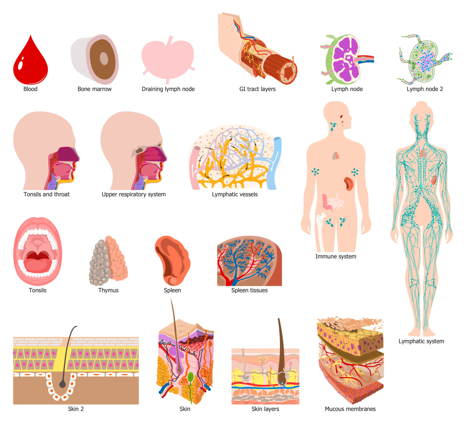
This sample diagram shows the organs of the human immune system. The immune system is a complex of cells, proteins, tissues and organs that are responsible for protecting the body from various infections, microorganisms, microbes and other harmful substances. The immune system has a vital role and is like a large accounting system that keeps a record of each germ, which ever entered in an organism and was defeated. And in a case of re-entering quickly recognizes and destroys these germs activating the adaptive immunity and innate immunity at the fight with invaders. The immune system is also involved in homeostasis and prevents an organism from the disrupting of normal functions by pathogens, that's why the immune booster is a key issue. As for the organs, which the immune system includes, the key among them are thymus and bone marrow, the secondary lymphatic organs are lymph nodes, lymph vessels, adenoids, tonsils, spleen, Peyer’s patches, and appendix.
Example 13: Vaccine Development
This diagram was created in ConceptDraw DIAGRAM using the combination of libraries from the Immunology Solution. An experienced user spent 10 minutes creating this sample.
This sample diagram lists various approaches used for HIV vaccine development. Many scientists are working on development an effective therapeutic vaccine to treat HIV-infected individuals and also preventive vaccine to protect people from being infected with HIV. Many different projects have been developed and a large number of researches in this area have been conducted over the past decades and the scientists' work doesn't stop today. It is an incredibly actual issue because a lot of deaths caused by HIV are fixed annually. The complexity of HIV vaccine development lies in a virus's high mutability and HIV isolates high variability. Among the main approaches are named whole inactivated, live attenuated, synthetic peptide, recombinant viral vector, recombinant bacterial vector, recombinant subunit, and plasmid DNA. The professionally designed objects from the solution libraries are the best way to make your immunology and medical diagrams illustrative and attractive.
Inside

What I Need to Get Started
After ConceptDraw DIAGRAM is installed, the Immunology solution can be purchased either from the Health area of ConceptDraw STORE itself or from our online store. Thus, you will be able to use the Immunology solution straight after.

How to install
First of all, make sure that both ConceptDraw STORE and ConceptDraw DIAGRAM applications are downloaded and installed on your computer. Next, install the Immunology solution from the ConceptDraw STORE to use it in the ConceptDraw DIAGRAM application.

Start using
Start using the Immunology solution to make the professionally looking illustrations by adding the design elements taken from the stencil libraries and editing the pre-made examples that can be found there.
