Medical Imaging
Medical imaging is one of the widely-used techniques of creating visual representations of the interior of a body. It may be used for either clinical analysis or medical intervention, or as a visual representation of the function of some tissues or organs. Medical imaging is widely used for revealing the internal structures hidden by the bones and skin. It also helps to diagnose and to treat the diseases. Medical imaging also can be used for establishing a database of normal anatomy, enabling to identify abnormalities.
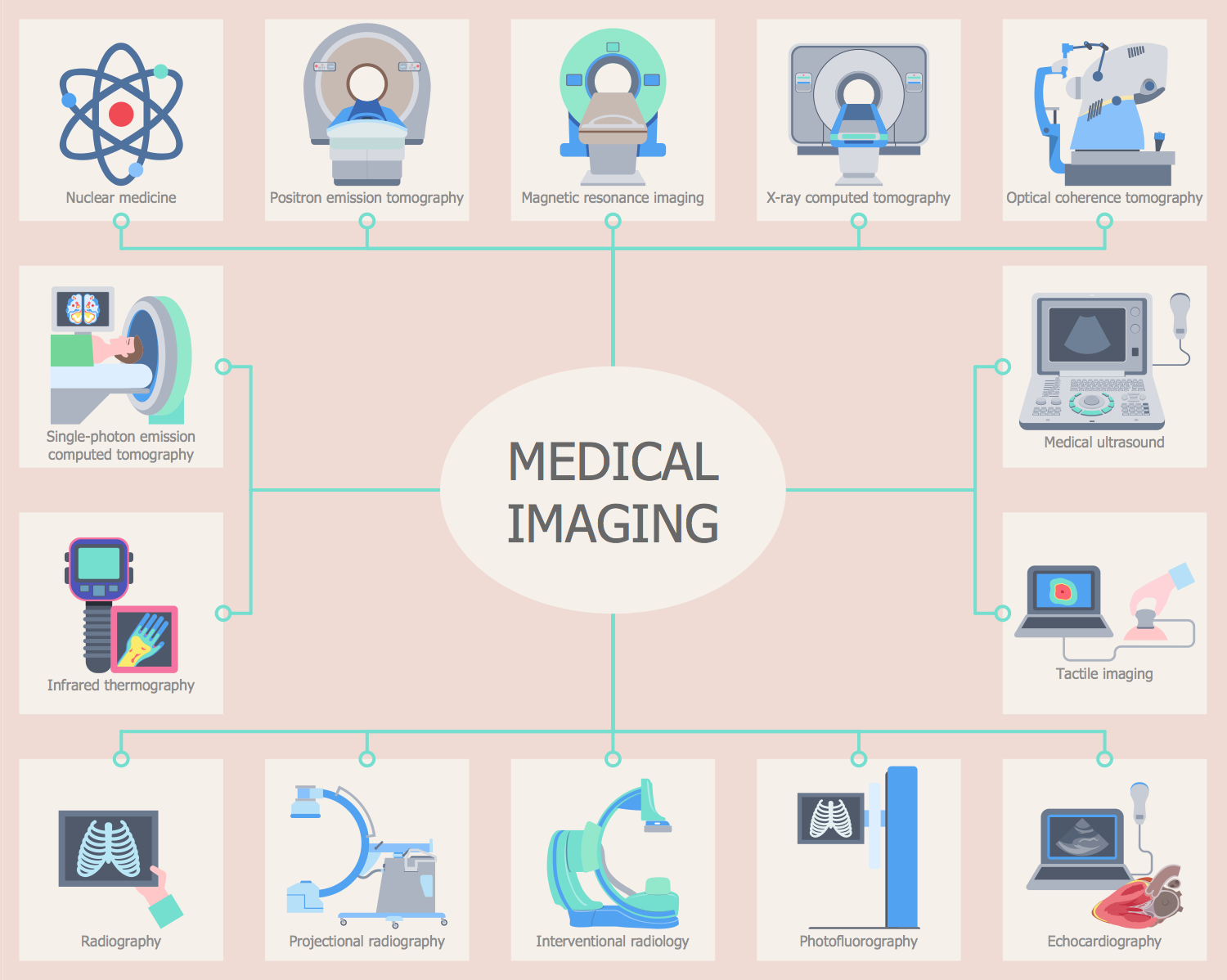
As a discipline, medical imaging is known to be a part of biological imaging, incorporating radiology which uses the imaging technologies of magnetic resonance imaging, X-ray radiography, medical ultrasonography (ultrasound), elastography, endoscopy, tactile imaging, thermography, medical photography, nuclear medicine functional imaging techniques, such as Single-photon emission computed tomography and positron emission tomography.
Recording and measurement techniques, such as electroencephalography, magnetoencephalography, electrocardiography, etc., represent other technologies which produce data susceptible to representation as a parameter graph vs. time or maps, containing data about the measurement locations. These technologies can be also considered as forms of medical imaging in another discipline.
Up to 5 billion medical imaging studies had been conducted worldwide until 2010. Being often perceived as a tool for designating the techniques that produce images of the internal aspect of the body, medical imaging can be a solution of numeral mathematical inverse problems. Thus, in the case of medical ultrasonography, the probe stands for ultrasonic echoes and pressure waves that go inside the tissue to show some internal structure. In the case of projectional radiography, the probe uses X-ray radiation that is known to be taken in by different tissue types in different amounts (e.g., bone, fat, and muscle).
Medical Imaging in ConceptDraw DIAGRAM
In the clinical context, the so-called "invisible light" medical imaging is usually equated to radiology or another term - "clinical imaging". The medical practitioner responsible for interpreting the images is called as a radiologist.
The so-called "visible light" medical imaging involves either digital video or still pictures which can be seen when you have no special equipment. Wound care and dermatology are two modalities that are known to be using visible light imagery. Diagnostic radiography is what designates the technical aspects of all medical imaging as well as the acquisition of medical images, in particular. The radiologic technologist (also known as “radiographer”) is responsible for acquiring medical images of diagnostic quality.
Medical imaging constitutes a sub-discipline of biomedical engineering, medicine or medical physics depending on the context. Thus, it may be used for development and research in the area of instrumentation, image acquisition, modelling and quantification are usually the preserve of biomedical engineering, computer science, and medical physics. It also can be used as a research into the interpretation and application of medical images is the preserve of radiology as well as the medical sub-discipline which is relevant to medical condition/area of medical science (cardiology, neuroscience, psychology, psychiatry, etc.) under investigation.

Medical Imaging using ConceptDraw DIAGRAM
Tactile imaging is another kind of medical imaging modality. It is used for translating the sense of touch into a digital image. It closely mimics manual palpation, deforming soft tissue by the probe and detecting all the resulting changes in the final pressure pattern.
Photoacoustic imaging is known to be a recently developed hybrid biomedical imaging modality. Photoacoustic imaging is based on the photoacoustic effect. The photoacoustic imaging can be also utilized in vivo for monitoring of tumour angiogenesis along with functional brain imaging, skin melanoma detection, and blood oxygenation mapping.
Echocardiography is next medical imaging technique described in this article. It allows one to see the detailed structures of the heart, such as heart function, chamber size, the valves of the heart and the pericardium. Echocardiography uses 3D, 2D as well as the so-called “Doppler” imaging in order to create pictures of the heart, visualizing the blood flowing through each of the heart valves. Echocardiography can be used for the patients suffering from the experiencing symptoms, such as chest pain, shortness of breath, etc.
Transthoracic ultrasound is known to be relatively safe for all patients of all ages. Thus, there is no risk of radiation or harmful side effects, comparing it to some other imaging modalities.
Echocardiography is known to be one of the most commonly used imaging modalities in the world. It is that popular because of its use and portability in many applications. Echocardiography is easily accessible and quick, allowing being performed at the bedside, which is what people need in emergency situations. The mentioned benefits of using echocardiography make it the modality of choice for many physicians.
In order to make the needed medical drawing, such as describing the medical imaging results, the ConceptDraw DIAGRAM diagramming and drawing software can be used. All the needed tools for creating the smart-looking as well as the professionally-looking drawings are there in the mentioned product of CS Odessa. In case the medical imaging-related drawings have to be created, then the Medical Illustrations solution may be also used.

Medical Imaging using ConceptDraw DIAGRAM
Offering the stencil libraries as well as the pre-made templates and examples of the medical illustrations, the Medical Illustrations solution may be chosen in order to simplify the work of creating the medicine-related drawings. Having the mentioned tools, any ConceptDraw DIAGRAM user can draw a professionally-looking medical illustration based on the already created templates.